VITA Zahnfabrik
H. Rauter GmbH & Co. KG
Spitalgasse 3
79713 Bad Säckingen

Prosthetic composite solution of ZrO2 and hybrid ceramic for a high chewing load
In the case of parafunctions, manifested bruxism and implant-supported dentures, prosthetic restorations are exposed to particularly high loads. Due to the enormous chewing forces, the risk of fractures or chipping increases in such cases. Prosthetic composite solutions can minimize these risks. In their case study, master dental technician Hans Jürgen Lange and dentist Dr. Michael Weyhrauch profile the restoration of a patient using composite bridges. This restoration concept is based on a high-strength zirconia framework structure and an elastic hybrid ceramic veneering structure.
1. The assessment
A 52-year-old woman suffered from temporomandibular joint pain and showed clear signs of bruxism on the tooth substance. Despite successful splint therapy, a new all-ceramic bridge restoration was fractured from 43 and 44 to 47 in the fourth quadrant. Even a longterm temporary restoration made of PMMA could not withstand the increased chewing forces for long. The dentist and dental technician discussed the case and decided to provide the patient with composite bridges made of VITA YZ T zirconia and VITA ENAMIC multi-Color hybrid ceramic (both VITA Zahnfabrik, Bad Säckingen, Germany).
2. The composite concept
With flexural strengths of around 1200 MPa, zirconia has proven to be extremely effective as a high-strength framework material. However, under extreme chewing force, fractures or chipping can occur, especially in the area of veneering, since all-ceramics have a high brittleness. Elastic materials with chewing force-absorbing properties like the hybrid ceramic VITA ENAMIC, are an interesting material alternative to consider here. In a composite bridge, the high strength of a zirconia framework structure is intelligently combined with the elasticity of a hybrid ceramic veneering structure. The hybrid ceramic VITA ENAMIC is based on a structure-sintered glass ceramic matrix (86% by weight), which is then infiltrated with a polymer (14% by weight). Due to this unique dual ceramic-polymer network structure, the material has a dentin-like elasticity, which can be expected to have a positive effect on restorations with a high chewing force load.
3. Digital workflow
Impressions of the bridge abutments were taken analogously to make the composite bridge. Based on this, a master model was made and digitized with the inEos X5 laboratory scanner (Dentsply Sirona, Bensheim, Germany). On the virtual model, a fully anatomical bridge was first designed using the exocad software (exocad, Darmstadt, Germany), which was then anatomically reduced at the press of a button. The framework was milled, reworked, sintered and scanned again to construct six monolithic veneering structures on top and then also produced with CAD/CAM support using the inLab MC XL System (Dentsply Sirona, Bensheim, Germany).
4. Finalization and integration
The hybrid-ceramic veneer structures were etched with hydrofluoric acid at the bonding surfaces and silanized, and the zirconia substructure was sandblasted. Adhesive bonding was performed with the dual-curing composite cement RelyX Unicem 2 Automix (3M, Seefeld, Germany). After the removal of composite residues, the final polishing was done with a goat hair brush and diamond polishing paste. Since 2017, when self-adhesive was integrated, the composite bridges have been complication-free in situ. The patient was enthusiastic about the pleasant chewing sensation, similar to that of natural teeth.
Report 07/19
Dr. Michael Weyhrauch, Mühltal, and Hans Jürgen Lange, Darmstadt, Germany
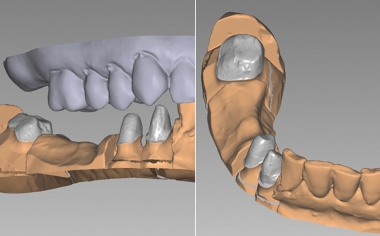
Fig. 1: Initial Situation with prepared stumps at 43, 44 and 47.
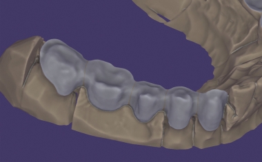
Fig. 2: The anatomically reduced bridge framework in the exocad software.
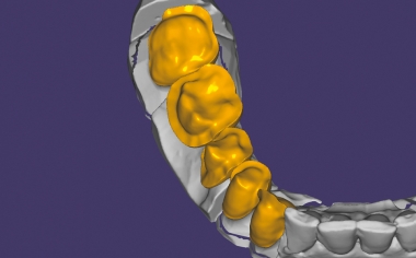
Fig. 3: Veneering structures made of hybrid ceramic were constructed on the zirconia framework produced with CAD/CAM support.
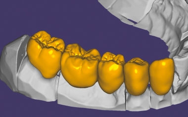
Fig. 4: Thanks to the low minimum wall thicknesses of the hybrid ceramic of up to 0.2 mm, the morphology is very natural.
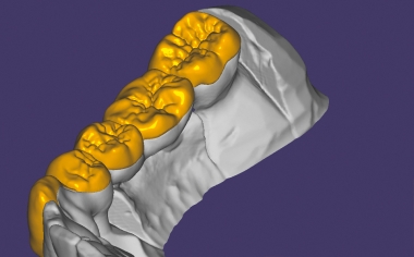
Fig. 5: The veneering structures in the equatorial area of the anatomically reduced framework construction end in the palatal area.
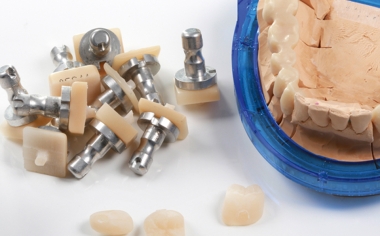
Fig. 6: Within one hour, the veneering structures were ground with the inLab MC XL unit.
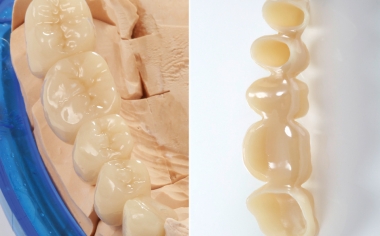
Fig. 7: The completely bonded bridge construction on the model from the occlusal and lumen side.
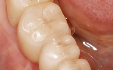
Fig. 8: The final integrated composite bridge from the occlusal side.
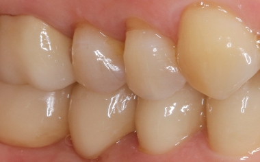
Fig. 9: Result: Intraorally, the bridge construction is functionally and esthetically very well integrated.