
Endocrown restoration made of proven VITABLOCS feldspar ceramic
A full crown preparation of deeply damaged teeth often results in the loss of large parts of the tooth walls, and, as a result, leads to further weakening of the tooth substance, as well as loss of retention. For the greatest possible preservation of natural tooth substance, a defect-oriented procedure using endocrowns is therefore recommended. In this case documentation, the VITABLOCS Mark II feldspar ceramic (VITA Zahnfabrik, Bad Säckingen, Germany) was processed for the time-saving and economic production of such a crown. The world’s first CAD/CAM material has proven itself millions of times since its first clinical use over 30 years ago. Clinical studies show a survival rate of 99.6% for endocrown restorations made of feldspar ceramic, after an observation period of seven years. Dr. Oxana Naidyonova explains her process below.
1. Assessment and pretreatment
A 48-year-old female patient came to the practice because tooth 34 was fractured and had previously been classified as not worth preserving by another practitioner. The clinical inspection showed an extensive disto-oral defect. The gingival tissue had overgrown into the cavity. X-rays showed an insufficient root canal filling. Since a full-crown preparation would have resulted in a loss of the vestibular and mesial wall portions of the tooth, the practitioner opted for an endocrown made of VITABLOCS Mark II. Tooth 34 was constructed with composite after a gingivectomy with laser, and a revision treatment was carried out.
2. Preparation and intraoral scan
Before the preparation, the tooth shade 2M2 was determined using the VITA Toothguide 3D-MASTER (VITA Zahnfabrik, Bad Säckingen, Germany), and the appropriate blank was selected. A glass fiber pin was adhesively introduced for additional retention of the subsequent composite structure. During the preparation, the walls were merely shortened and a groove was created in the defect area. Sharp edges in the cavity were consistently rounded off. Prior to the intraoral scan, the proximal caries on tooth 34 could be mesially and minimally invasively treated with composite, thanks to its good accessibility.
3. CAM and finalization
The endocrown was then digitally designed and fabricated from VITABLOCS Mark II with the My-Crown Mill grinding unit (FONA Dental, Bratislava, Slovakia). After separation from the attachment, the restoration was tried in and then gently finalized with a fine diamond. This was followed by the characterization of the fissures with VITA AKZENT Plus EFFECT STAINS (ES06, rust red) and the final glaze. Since a reliable adhesive bond to the tooth substance is a key component of long-term clinical success, a rubber dam was created to ensure freedom from contamination and absolute dryness.
4. Conditioning and integration
The feldspar ceramic was then etched with hydrofluoric acid to create a microretentive etch pattern and then silanized. The cavity was conditioned with phosphoric acid and an adhesive. For adhesive bonding, the composite Micerium (Micerium, Avegno, Italy) in the shade UD2 was heated to make it flowable for insertion. After light-curing and removal of composite residues, the restoration was well integrated into the natural tooth structure, thanks to its outstanding light-optical properties.
Report 07/19
Dr. Oxana Naidyonova, Karaganda, Kazakhstan
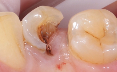
Fig. 1: Initial situation: Tooth 34 was severely damaged. Gingival tissue had overgrown into the cavity.
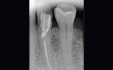
Fig. 2: Status following revision, pin setting and build-up filling.
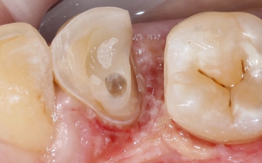
Fig. 3: Care was taken during the preparation not to leave any sharp edges in the cavity.
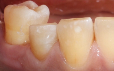
Fig. 4: The remaining cavity walls were shortened only occlusally.
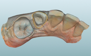
Fig. 5: After the intraoral scan, the preparation margin was determined.
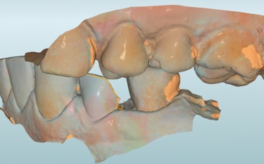
Fig. 6: The habitual intercuspation was transferred with a vestibular scan.
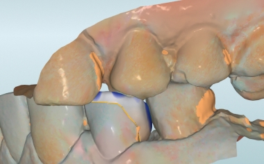
Fig. 7: The minimum layer thicknesses were maintained in the construction of the restoration.
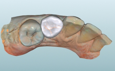
Fig. 8: The virtual endocrown in the CAD software from the occlusal view.
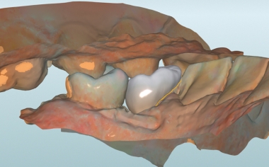
Fig. 9: The construction in the lingual view.
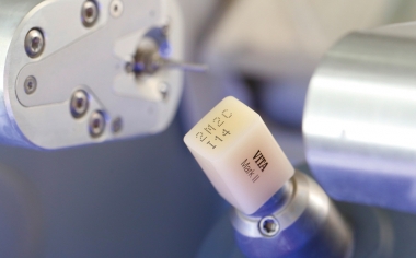
Fig. 10: The VITABLOCS Mark II clamped in the grinding unit.
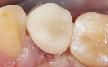
Fig. 11: The feldspar ceramic restoration during the clinical try-in.
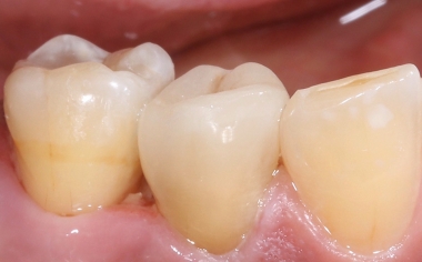
Fig. 12: No transition between the restoration and the tooth can be seen from the vestibular view.
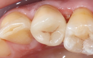
Fig. 13: The occlusal view of the fully adhesively integrated endocrown.
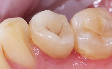
Fig. 14: Result: The highly esthetic integration of the restoration at the time of follow-up after six months.