
Minimally-invasive restoration of an incisal edge defect with CAD/CAM hybrid ceramic
The worldwide unique CAD/CAM hybrid ceramic VITA ENAMIC consists of a structure-sintered glass ceramic matrix that is infiltrated with polymer. The dual ceramic-polymer network allows for very delicate reconstructions with wafer-thin, precise marginal areas of up to 0.2 millimeters. Thanks to its dentin-like elasticity, its enamel-like abrasion behavior and its natural light transmission, the CAD/CAM material exhibits excellent functional and esthetic integration into the natural tooth structure. In the following case study, Dentist Dr. Sheng Fang (Chengdu, China) and Dental Technician Feng Li (Chengdu, China) show how they were able to restore an incisal edge defect with a minimally invasively process on the central maxillary anterior tooth using the hybrid ceramic VITA ENAMIC (VITA Zahnfabrik, Bad Säckingen, Germany).
1. The patient case
A 21-year old patient visited the dentist's office because the composite structure of her distal corner of tooth 21 was fractured due to secondary caries. She wanted a long-term, permanent restoration to be integrated harmoniously into her tooth structure. Because this restoration was scheduled to be minimally invasive and required a reconstruction with thin walls, the dentist and dental care team opted for a CAD/CAM-supported reconstruction using the hybrid ceramic VITA ENAMIC.
2. Tooth shade determination and material selection
Precise color information is essential in making the correct choice when it comes to the shadematching material blank. To ensure optimal shade integration of the existing incisal edge defect on the reconstruction, the tooth shade was determined using the VITA Linearguide 3D-MASTER shade guide, after applying local anesthesia. Systematic tooth shade determination was carried out in two steps. In the first step, a brightness level from 0 to 5 was determined using the VITA Valueguide 3D-MASTER shade guide. The color intensity and shade were then determined using the appropriate shade guide from the VITA Chroma/ Hueguide 3D-MASTER. This resulted in the selection of tooth shade 1M2. Since this primarily involved a restoration of the translucent enamel area, a translucent HT blank in the color 1M2 was selected for the CAD/CAM-supported fabrication. To prepare for the digital impression, only the caries was removed and the enamel edges of the defect were slightly tapered.
3. CAD/CAM fabrication and finishing
This was followed by the intraoral scan with the CEREC Omnicam 4.2 and the virtual design of the restoration using the CAD software inLab CAD 15.2. The order was sent to the inLab MC XL milling unit and executed there (all Dentsply Sirona, Bensheim, Germany). Afterwards, the sprue was removed and the restoration was finished with fine diamond instruments. Lastly, the final finishing was done using the VITA ENAMIC Polishing Set technical. During the try-in, the partial restoration was a perfect fit and could be etched on the adhesive surfaces using hydrofluoric acid and then silanized. The tooth substance was pre-treated using the acid etching technique and then an adhesive was applied. This was followed by final seating using composite cement.
4. Finalization and summary
After the cement residues were removed, the transitions between tooth and restoration were evened out using the VITA ENAMIC Polishing Set. The delicate restoration showed a very harmonious integration in the natural tooth structure, thanks to its natural play of colors and light. Due to the comparatively low brittleness of hybrid ceramic - even with very thin wall thicknesses - and its thinly tapered edges, hybrid ceramic can be precisely processed and the patient provided with minimally invasive treatment. Using the digital workflow to create an efficient fabrication of the indirect restoration made it possible to treat the patient in one session. The dental team and the patient were completely satisfied with the results of the final restoration.
Report 04/20
Dentist Dr. Sheng Fang Chengdu, China and
Dental Technician Feng Li Chengdu, China
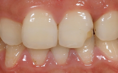
INITIAL SITUATION The initial situation with fractured tooth 21 when the patient first presented at the dental practice.
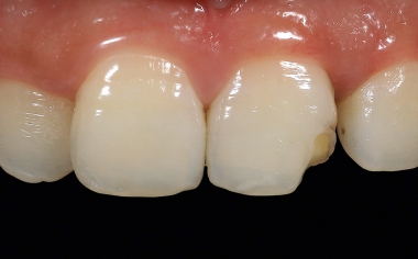
Fig. 2 Secondary caries had formed under a direct composite abutment, which led to a filling fracture.
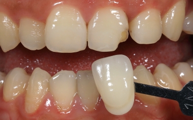
Fig. 3 The tooth shade was systematically determined in two steps using the VITA Linearguide 3D-MASTER.
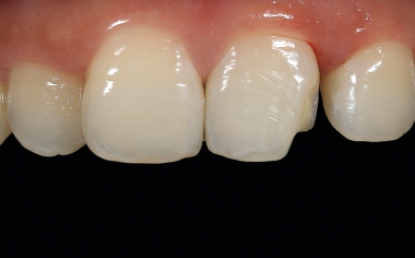
Fig. 4 The caries was removed under local anesthesia and the edge areas in the enamel were slightly tapered.
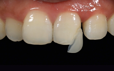
Fig. 5 The CAD/CAM-supported finished restoration made of VITA ENAMIC with wafer-thin edges.
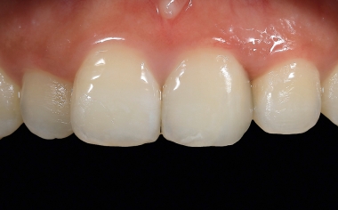
RESULT The final situation after the fully adhesive integration with composite.