
Non-invasive microveneer restoration made of the hybrid ceramic VITA ENAMIC
Producing noninvasive microveneers using CAD/CAM systems has been a major challenge so far, due to the brittleness of ceramic dental materials. The very small wall thicknesses and thinly tapering edge areas of such restorations often showed significant ceramic chipping or fractures after CAM production. The dental team of Dr. Michael Tsao and Dr. Hsuan Chen systematically examined a variety of ceramic material samples in numerous test runs. They selected the hybrid ceramic VITA ENAMIC (VITA Zahnfabrik, Bad Säckingen, Germany) for their restoration. Based on their clinical experience, this material still allows a very good marginal integrity even with wall thicknesses of 0.2 mm. In this report, the practitioners show the noninvasive, model-free and fully digital restoration of a diastema.
1. Assessment and planning
A 29-year-old male patient presented in the practice because he was dissatisfied with his diastema between teeth 11 and 21. Orthodontic therapy was rejected by the patient. He wanted a time-efficient solution with the greatest possible preservation of the natural tooth substance. The usual production of microveneers on refractory stumps was too lengthy for him. Therefore, the practitioners and patient decided to implement the gap closure in the digital workflow using VITA ENAMIC hybrid ceramics in a single session.
2. Tooth shade determination and digital design
After meticulous cleaning of the restoration area, the tooth shade was determined with the VITA Toothguide 3D-MASTER (VITA Zahnfabrik, Bad Säckingen, Germany) on the two central incisors in the upper jaw. The tooth shade 1M2 was determined and the corresponding blank selected. After retraction threads were laid, the intraoral scan was performed with CEREC Omnicam (Dentsply Sirona, Bensheim, Germany). Because of the high enamel translucency of the tooth in the approximal region, scanning powder was applied to facilitate intraoral scanning. Sirona Connect transferred the data record to the inLab software. There, the razor-thin microveneers were digitally constructed.
3. CAM fabrication with a highly precise result
For CAM production with the CEREC MC XL System (Dentsply Sirona, Bensheim, Germany), the VITA ENAMIC blanks were fixed in the grinding unit and the corresponding grinding job was carried out. The grinding result showed razor-thin microveneers with absolutely precise edge areas. Thanks to its dual ceramic-polymer network structure, the hybrid ceramic has a significantly higher elasticity and less brittleness than traditional ceramic CAD/CAM materials. This enables high-precision reconstructions with simultaneously low wall thicknesses. Finally, the graceful restorations were carefully separated from the attachment with a fine diamond, finalized and tried-in.
4. Conditioning according to proven protocol
VITA ENAMIC has a stable, highly networked ceramic matrix. The ceramic portion of the material is 86 percent (wt%). As a result, the hybrid ceramic can be preconditioned using hydrofluoric acid and silane, according to the traditional, all-ceramic protocol. CAD/CAM composites, on the other hand, are sandblasted because they have a polymer matrix in which ceramic fillers are embedded. Sandblasting, however, can lead to damage of the material structure and thin edge areas in reconstructions with low wall thicknesses. In the present case, the microveneers were pretreated with the safer protocol. The enamel was conditioned with phosphoric acid and adhesive. Afterwards, the veneers were fixed with a composite cement. After removal of the composite residues and polishing with the VITA ENAMIC Polishing Set, a highly esthetic result was achieved efficiently and noninvasively after only one session.
Report 7/19
Dr. Michael Tsao and Dr. Hsuan Chen, CEREC Asia, Taipeh, Taiwan
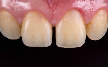
Fig. 1: Initial sizuation: Young patient with diastema between teeth 11 and 21.
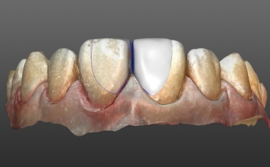
Fig. 2: Razor-thin veneers were constructed using inLab software.
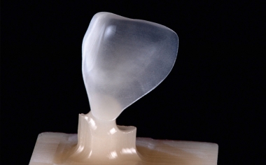
Fig. 3: The high-precision result after the grinding process with CEREC MC XL.
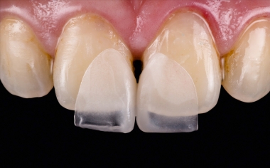
Fig. 4: The delicate microveneers on tooth 11 and 21 during the clinical try-in.
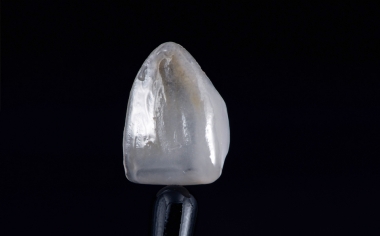
Fig. 5: Veneer characterized with light-curing stains before seating.
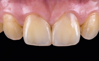
Fig. 6: There was a natural light transmission following the fully adhesive attachment.
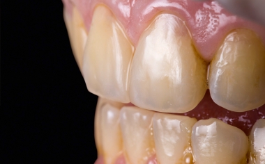
Fig. 7: A transition-free and natural appearance was also evident laterally.
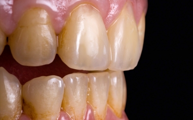
Fig. 8: Result: The patient was very satisfied with the highly esthetic result in only one session.